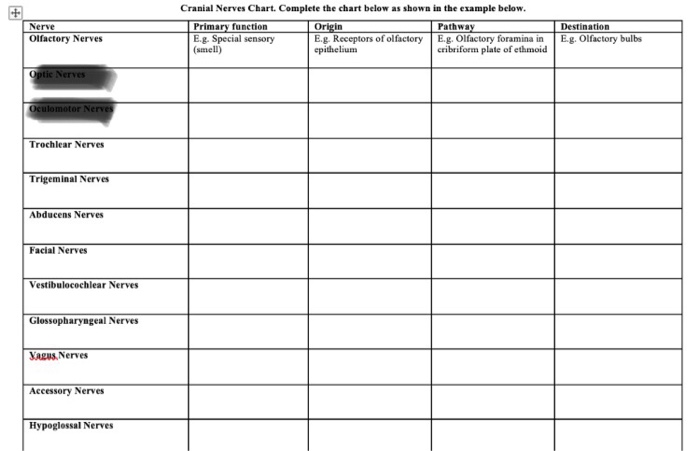
Cranial Nerves Function Chart
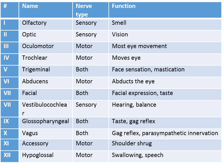
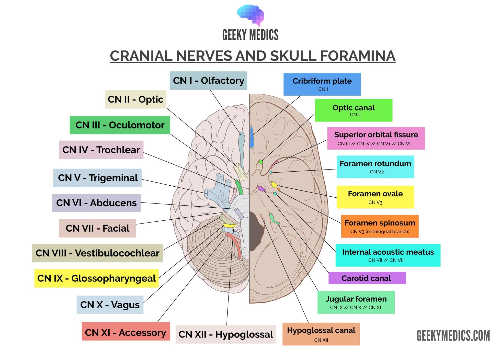
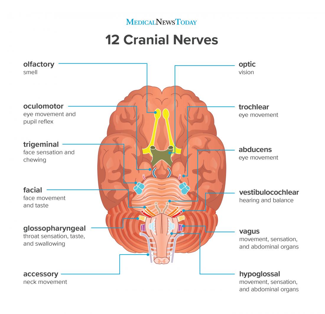
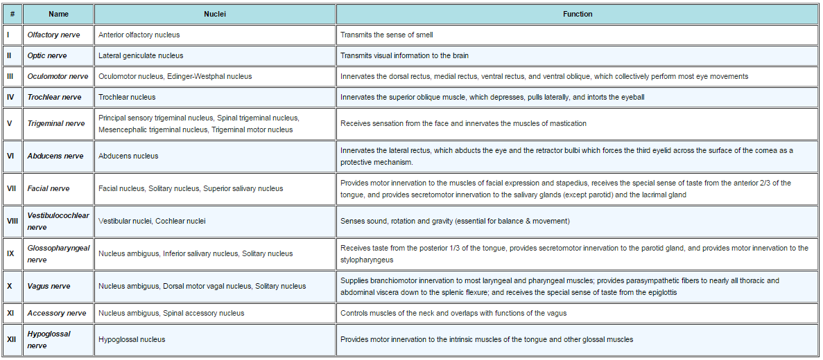
The cranial nerves are twelve pairs of nerves from the central nervous system.
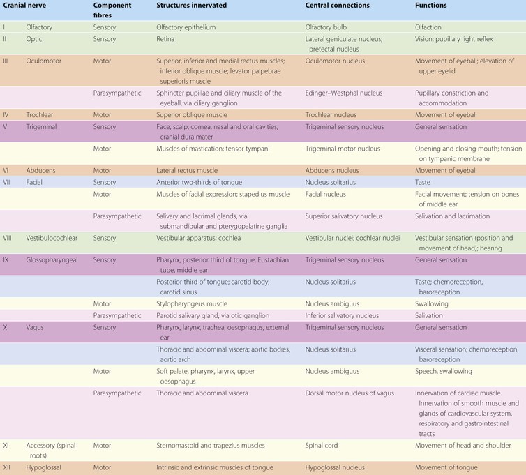
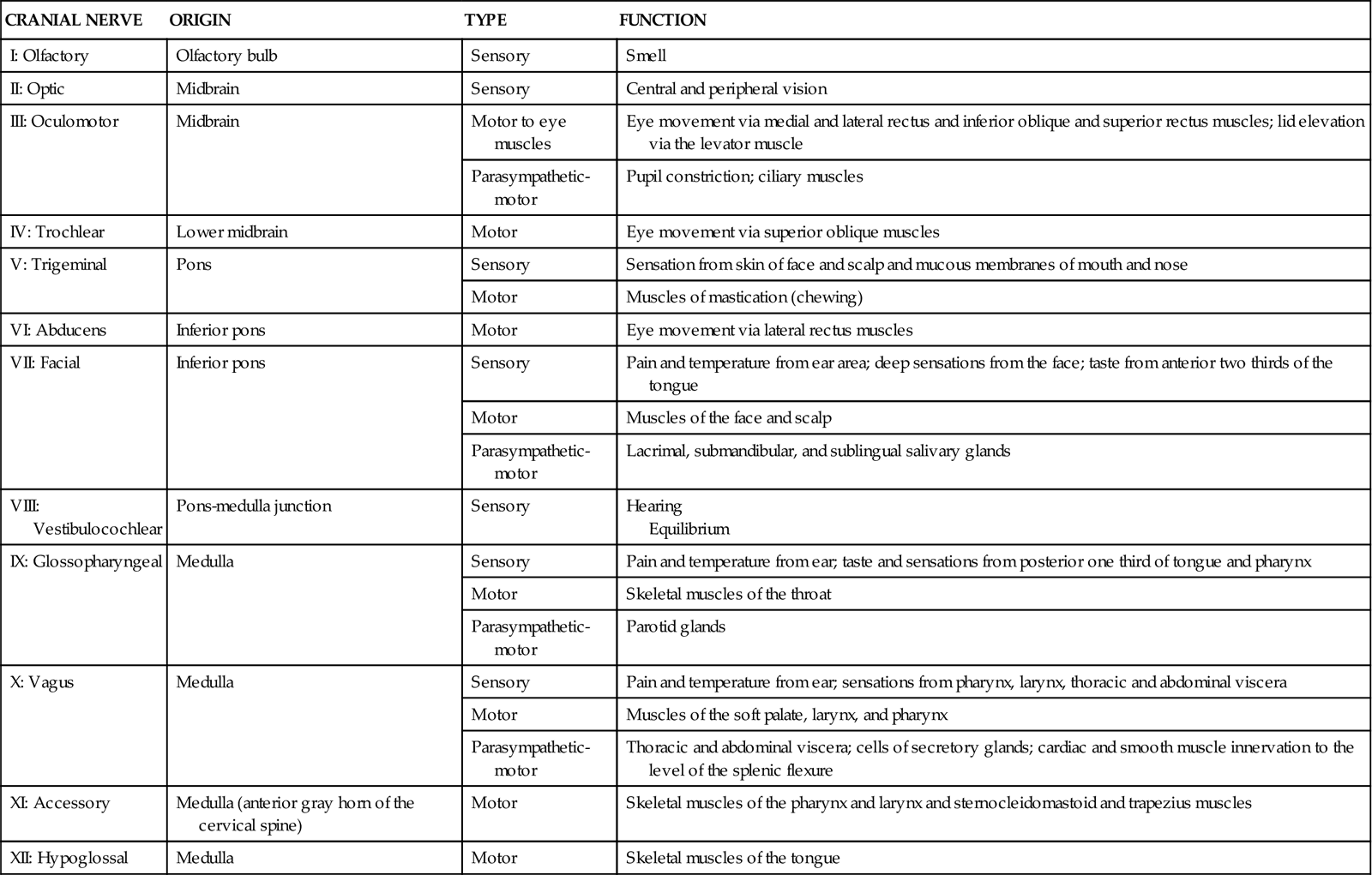
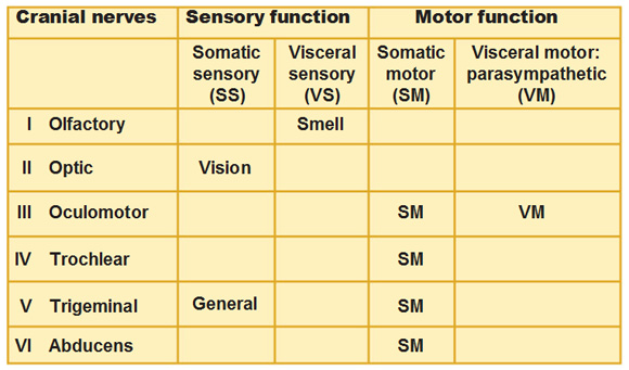
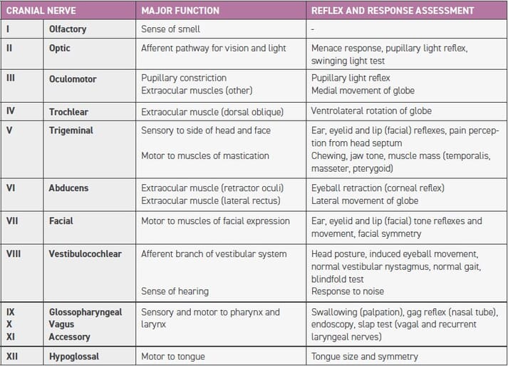
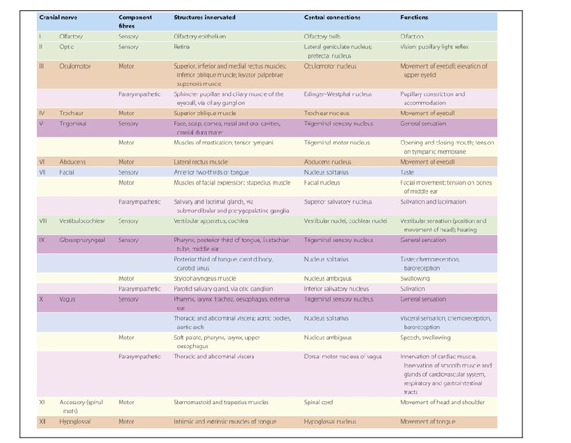
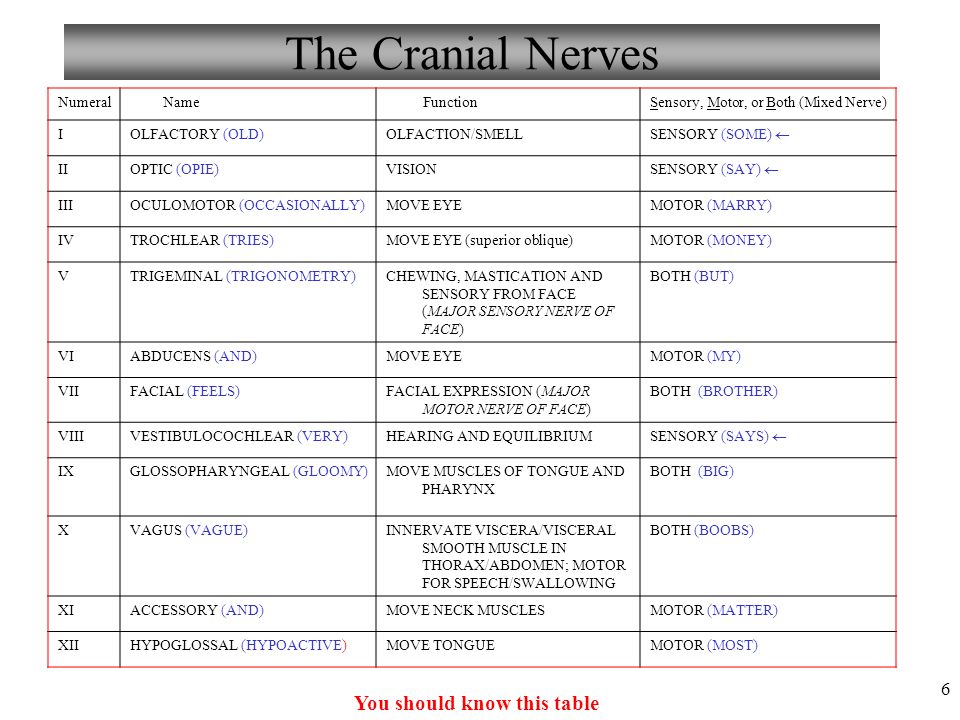
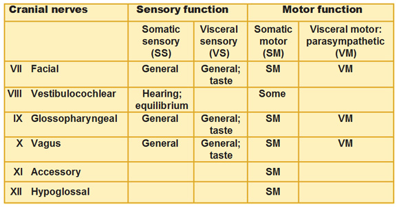
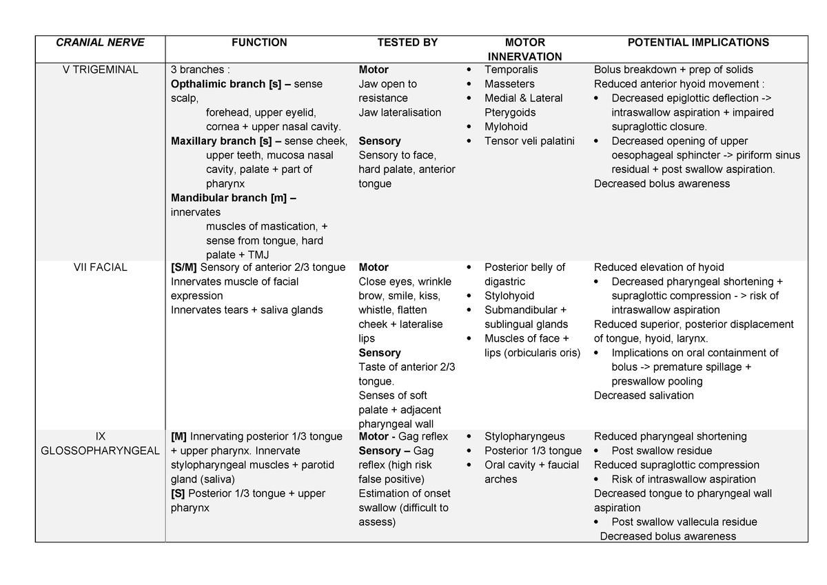
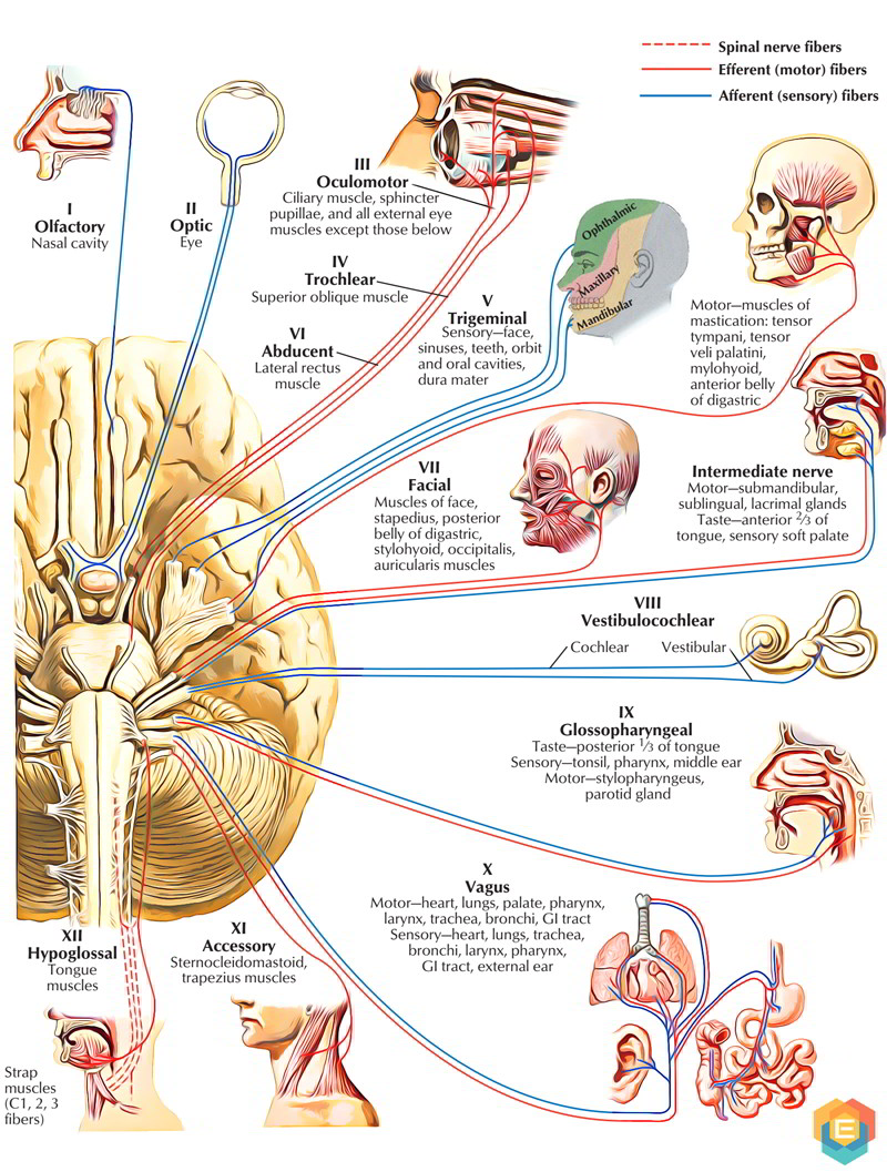
Cranial nerves function chart. Receives sensation from the face and innervates the muscles of mastication. V 1 ophthalmic nerve is located in the superior orbital fissure v 2 maxillary nerve is located in the foramen rotundum. The functions of the cranial nerves are sensory motor or both.
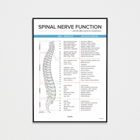
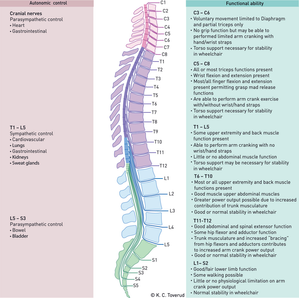
2 c1 does not have a dermatome. 1 the c2 dermatome handles sensation for the upper part of the head and the c3 dermatome covers the side of the face and back of the head. C1 c2 and c3 the first three cervical nerves help control the head and neck including movements forward backward and to the sides.
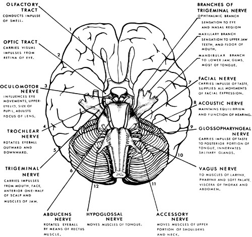
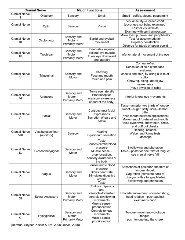
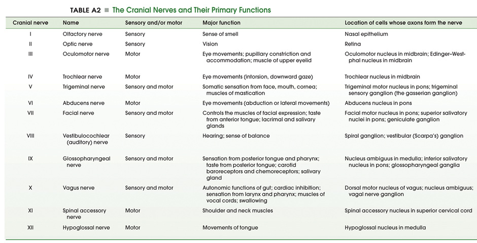
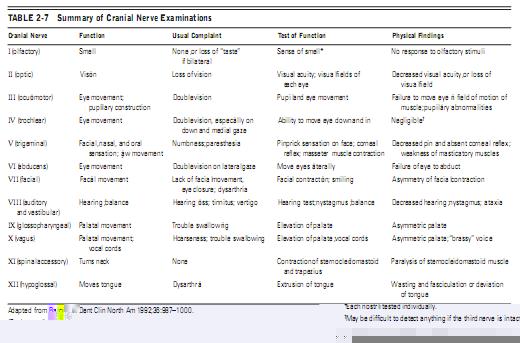
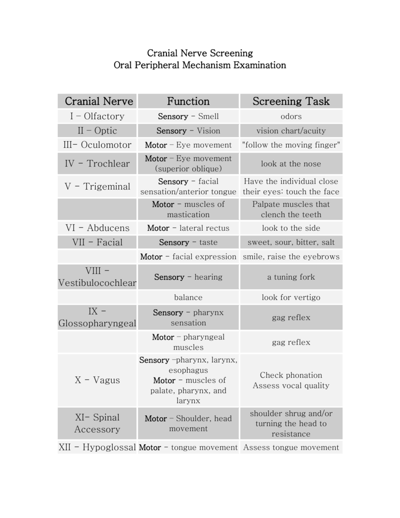
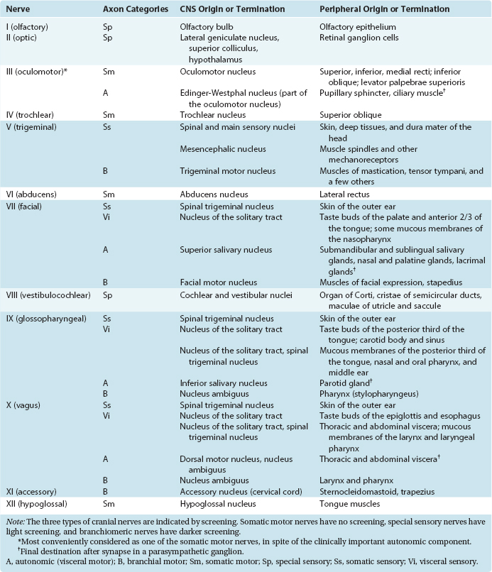
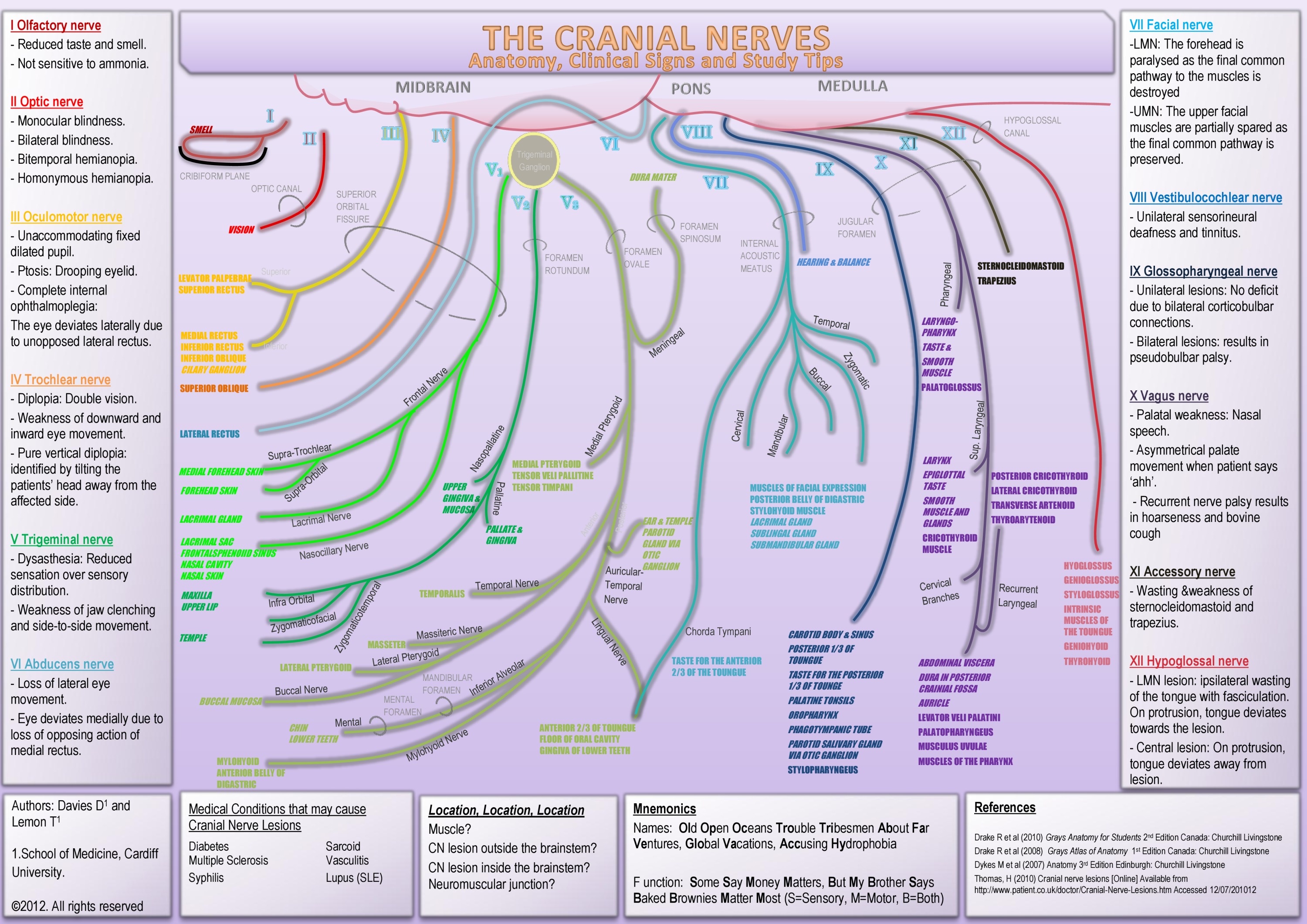
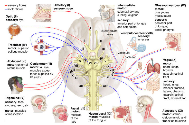
Cranial nerves are responsible for the control of a number of functions in the body. Some of these functions include directing sense and motor impulses equilibrium control eye movement and vision hearing respiration swallowing smelling facial sensation and tasting. Your cranial nerves are pairs of nerves that connect your brain to different parts of your head neck and trunk.
Each nerve also. List of the 12 cranial nerves with concise information about the name number and functions of each. This is part of the human anatomy pages about the nervous system.
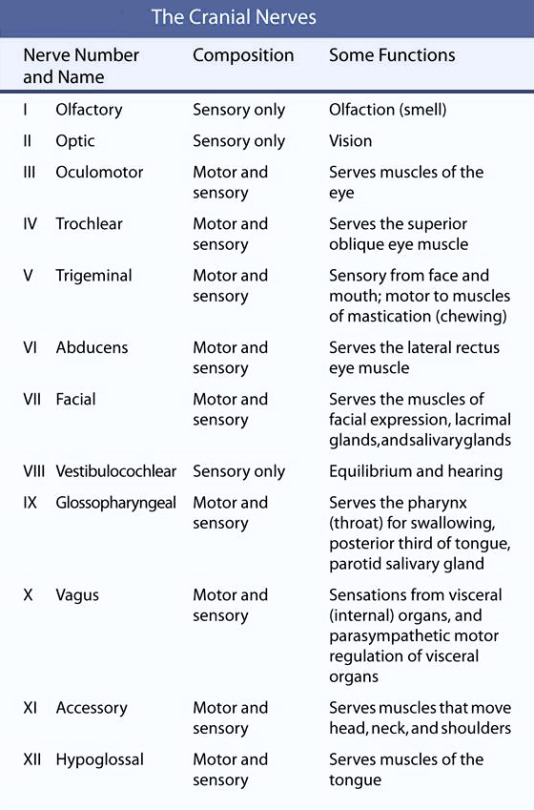
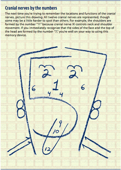
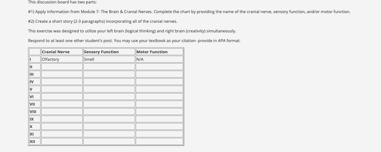
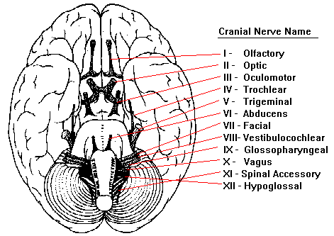
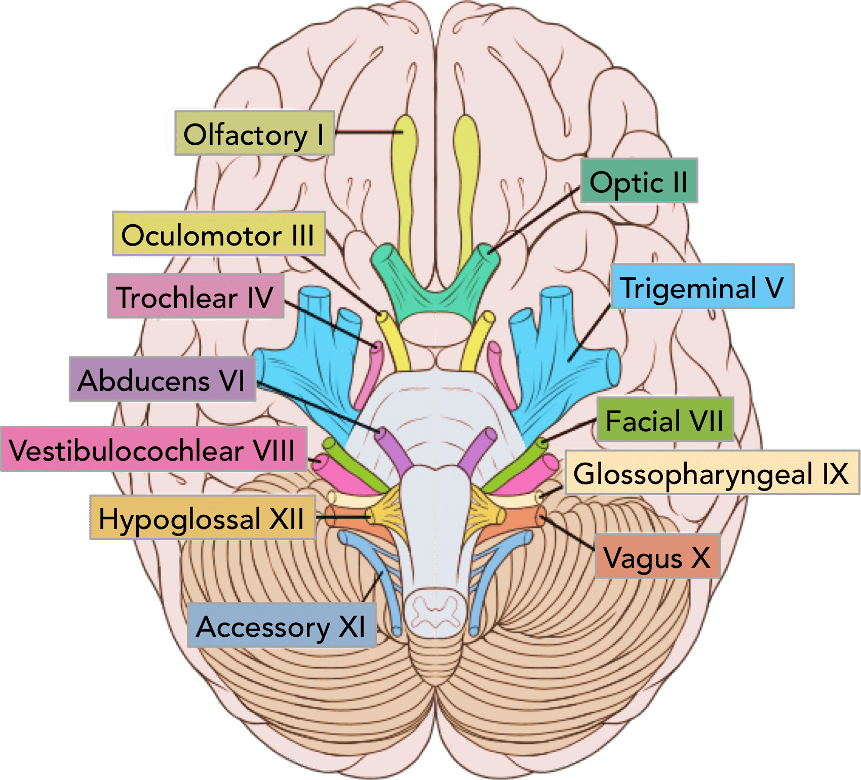
In this summary we discuss the nomenclature of the cranial nerves and supply some background information that might make it easier to understand the nerves and their function. The cranial nerves listed here are i olfactory ii optic iii oculomotor iv trochlear v trigeminal vi abducens vii facial viii vestibulocochlear ix glossopharyngeal x vagus xi accessory and xii hypoglossal. Motor cranial nerves help control muscle movements in the head and.
V 3 mandibular nerve is located in the foramen ovale. The major function of cranial nerves is to send and receive information from the brain to the various body parts. Cranial nerve major functions assessment.
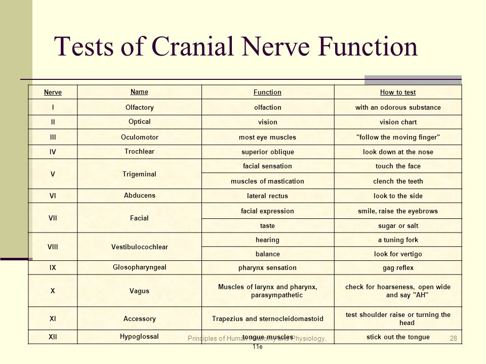
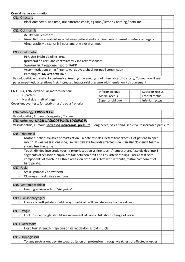
Cranial nerve assessment normal response documentation. The cranial nerves are loosely based on their functions. There are 12 of them each named for their function or structure.
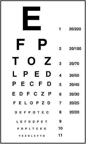
Cranial nerve i olfactory sensory smell smell coffee cloves peppermint cranial nerve ii optic sensory vision visual acuity snellen chart cover eye not being examined test for visual fields examine with ophthalmoscope cranial nerve iii oculomotor sensory and motor primarily motor eyelid and eyeball movement move eye up down and peripherally test for accommodation pupillary constriction observe for ptosis of upper eyelid cranial nerve iv. Ask the client to smell and identify the smell of cologne with each nostril separately and with the eyes closed. The names and major functions of these nerves are listed below.






/cranial-nerves-2-1e3d489c9104495dbcc609ea188af32d.jpg)
:background_color(FFFFFF):format(jpeg)/images/article/en/the-12-cranial-nerves/BJlRqDAfAoB4SnDOELIzQ_Cranial_Nerves_Draft_3.png)







/cranial-nerves-2-1e3d489c9104495dbcc609ea188af32d.jpg)
















:max_bytes(150000):strip_icc()/Snellen_chart-7d82f27667194a91959b5ef2682d0a19.jpg)



:background_color(FFFFFF):format(jpeg)/images/library/12658/nervous-system-breakdown.png)


